

As a nationally-recognized specialist in hip arthroscopy, Dr. Carreira regularly fields questions from patients about hip problems and, more specifically, hip arthroscopy. From time to time, Dr. Carreira will post new questions or updated answers to these hip arthroscopy FAQ. Dr. Carreira maintains his medical practice in Atlanta, Georgie.
Key Hip Arthroscopy FAQ (Frequently Asked Questions)
These hip arthroscopy FAQ are general in nature. To discuss your own medical condition, please make an appointment with Dr. Carreira.
Q: Who is a good candidate for hip arthroscopy?
A: The main reason to perform a hip arthroscopy is to treat hip pain in those patients who have failed nonoperative treatments and who do not have diffuse areas of articular damage (arthritis). It is important to note that hip arthroscopy is a surgical procedure that can be used to treat and correct a variety of hip problems.
The most common injury for which hip arthroscopy is a viable option is a labrum (labral) tear. Other joint problems which can be treated at the time of arthroscopy include ligamentum teres tears (the central ligament of the joint), synovitis (inflammation of the joint lining), loose bodies (free floating pieces of cartilage and/or bone), and femoroacetabular impingement (abnormal bone shape on the acetabular (cup) or femoral (ball) side of the joint.
The typical patient who may benefit from hip arthroscopy is suffering from hip pain and does NOT have a diagnosis of hip arthritis. Arthritis is defined as generalized or diffuse articular cartilage injury of a joint. Hip pain typically occurs in the front and/or the side of the joint, and may also be present in the buttock. Rarely hip joint pain occurs only in the buttock. Patients may experience pain with movement of the hip joint, including flexion, such as when tying shoes or getting into a car, or at rest, such as when sitting or trying to sleep. Patients typically experience increased pain with increased activity, either during the activity itself, or in the day or two following the activity.
The majority of patients, as noted in our MASH Study Group of over 8,000 patients, have gradual onset of pain without a history of trauma or injury. Patients generally have “normal” x-rays that do not show any signs of arthritis. On close review of these x-rays however, forms of femoroacetabular impingement (FAI) may exist. An MRI typically is performed to detect labral tears, which are a common cause of chronic pain in the hip joint.
Q: What factors might limit the success of a hip arthroscopy procedure?
A: The main limitation in the success of hip arthroscopy is the presence of arthritis, or articular cartilage damage. If the area of damage is small, a microfracture technique at the time of arthroscopy is oftentimes successful in repairing the damaged cartilage.
If there is diffuse (wider spread) articular cartilage damage, meaning that damage of the articular cartilage is throughout the joint, the best treatment is a joint replacement.
X-rays are the best way to determine the extent of articular cartilage damage.
Q: What are the main risks of hip arthroscopy?
A: Infection – The risk of infection is very low and is decreased with a sterile operating environment and antibiotics, as well as careful handling of the incision sites following surgery. Signs and symptoms of an infection are redness, pain, swelling and warmth around the incisions and non-clear drainage from the incision. These infections are typically superficial and require cleaning with soapy water and occasionally antibiotics.
DVT (deep vein thrombosis, blood clot) – The risk of developing a DVT is decreased by starting early motion including the CPM machine, ankle pumps, aspirin or other blood thinner. Early movement with walker/crutches (not lying in bed immobilized) help to prevent clots. Signs and symptoms of a blood clot are pain and swelling most commonly in the calf. The calf may also become red and warm to the touch.
Pain – We try to minimize pain as much as possible both in the short and long term, with a goal of little to no pain with full activities after a few months. Medication, ice, rest and therapy reduce post-operative pain. Following restrictions after surgery help to ensure adequate healing of the tissues. “Little old grandma or grandpa level of activity” in the first 6 weeks is recommended.
Numbness – With hip arthroscopy, there is a very small chance of post-operative numbness in the groin and genitalia region and lower extremities. You may also experience numbness of the upper outer portion of the thigh on the operative leg after surgery. The lateral femoral cutaneous nerve is close to the surgical area and it may be stretched or bruised during the procedure. Numbness very commonly resolves and Dr Carreira is not aware of any permanent nerve injuries in the patients that he has treated.
Q: Where can I learn more about hip arthroscopy?
A: The American Academy of Orthopedic Surgeons (AAOS) has an entire section of its website dedicated to providing detailed information about hip arthroscopy. While there are no hip arthroscopy FAQ on the AAOS site, it does contain plenty of good information, photos, and diagrams.
Q: What might a hip exam look and feel like?
A: In the video below, Dr. Dominic Carreira explains the process of walks through what a typical hip exam might entail for his patients.
Q: What are the options for labrum treatment in hip arthroscopy?
A: There are three different forms of labrum treatment that have been described.
- Debridement is the simplest form of treatment and it consists of simply removing damaged tissue, most typically with a shaver. In my practice, this is rarely performed, given that the results of debridement is less predictable. However, at the time of surgery, there are situations in which the labral tear itself is irreparable, that is it is damaged in a way that re-fixating it is not an option. In these situations, the majority of patients still do have a good result with surgery.
- Repair is associated with better results than debridement, and the development of labral repairs in hip arthroscopy has increased success rates and been part of the reason that hip arthrscopy has grown so much in the past decade. Anchors are placed into the bone to suture the labral tissue back to the bone from which is has become detached. Success rates with repairs are very high, including to return to high demand sports and activities.
- Reconstruction is performed with cadaver tissue, and is performed when the tissue is irreparable. For a more detailed overview of labrum reconstruction in hip arthroscropy, please see the answer to the question immediately below.
Dr Carreira may discuss these options with you depending on your condition. The MRI, X-Rays, physical examination, and any other past history of treatment may be helpful in formulating a plan that best suits you and helps to ensure an excellent result.
Please note that patient case reviews may be performed for out of town patients. Please follow the directions on the Request a Free X-Ray + MRI review page.
Q: How frequently does Dr. Carreira perform a Hip Labral Debridement versus a Labral Repair versus a Labral Reconstruction?
A: The frequency of reconstruction has increased a lot in recent years given the increased evidence to support its use. Currently, Dr. Carreira performs approximately 1% debridements, 80% repairs, and 19% reconstructions. For those patients who need revision hip arthroscopy because of persistent pain, the use of reconstruction is much more common than in years past.
Q: What is the role of labrum reconstruction in hip arthroscopy?
A: Reconstruction is performed with cadaver tissue, and is performed when the tissue is irreparable. Dr Carreira takes the approach which most expert hip arthroscopists nationally have taken, which is that labrum reconstruction should not be performed the first time that a patient undergoes hip arthroscopy, as the other forms of treatment are likely to be successful, especially repairs. There are circumstances when patients have not done well with a first hip arthrscopy, and when the labrum is deemed to be in poor condition, either because of complex tearing, extensive bruising, a small amount of tissue, or the tissue has been debrided away, and labrum reconstruction is indicated. Dr Carreira has over 10 years experience with the surgery and has published high success rates with this surgery. Patients who undergo reconstruction are usually older and are more likely to have associated articular cartilage injuries. The cadaver tissue is introduced into the joint and serves the same function as a regular labrum.
Dr. Carreira’s experience with allograft reconstruction was accepted for publication to the Journal of Hip Preservation Surgery. The important finding of Dr. Carreira’s study include the fact that reconstruction is, in fact, a good option in select patients. That said, there is still some controversy among medical professionals as to which are the best patients for each type of treatment.
In Dr. Carreira’s series of patients with reconstructions, the patients were generally found to have more degeneration of the joint, with higher rates of articular cartilage damage and treatment, including microfracture. In 2018, two other publications have also demonstrated success with reconstructions, both of which are well-done studies in reputable journals. Labral reconstruction during hip arthroscopy is a reasonable option in a small percentage of patients whose labrum is deficient or irreparable.
The selection of debridement, repair, or reconstruction is a focus of study for the Multicenter Arthroscopy Study of the Hip.
Q: What are the general milestones and limitations related to recovery from hip arthroscopy surgery?
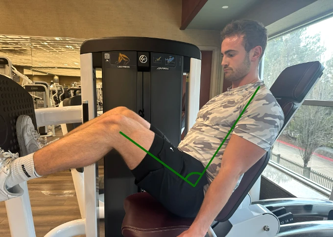
A man using the leg press machine while keeping the angle of his hips in a non-impinging position.
A: Simple activities of daily living are expected in the first 6 weeks after surgery. The phrase “like a little old grandma (or grandpa)” as a general guideline for levels of activity. This includes slow walking when moving about the house or getting dropped off in front of a restaurant as reasonable activities. We do not recommend fast walking, walking long distances for exercise, picking up heavy objects greater than 10 pounds, chasing the dog, etc.
After 6 weeks, normal daily living activities are reasonable.
High-level activities such as jogging, quick movements such as changing directions, and lifting heavy objects are all a minimum of 3 months after surgery. Based on the surgical findings, these guidelines may be adjusted to your particular situation.
Generally speaking, physical therapy will set the ceiling in terms of the highest amount of activities which are to be followed during the course of recovery. Pain is used as a guide in terms of progression. As the weeks pass, there is a general increase in activity and the progression is not only towards improvement. Some “bumps in the road” are expected.
Q: Why there are differences in interpretations of imaging studies across different providers?
A: These differences occur across different types of imaging studies, including x-rays, MRI’s, and occasionally, CT scans. There are many factors that contribute to these differences, including patient positioning, quality of the images themselves, the type of software using the machines, and the interpretation of the images by radiologists or orthopedic surgeons.
Dr. Carreira’s study and article, “The Reliability of Commonly Used Radiographic Parameters in the Evaluation of the Pre-Arthritic Hip: A Systematic Review”, recently was approved for publication. In the study, Dr. Carreira reviewed the reliability across different healthcare providers in the interpretation of plain x-rays. His study included interpretation of x-rays for the determination of arthritis as well as for different shapes of the hip including for cam and pincer impingement in femoroacetabular impingement (FAI) and for dysplasia.
Q: What is the risk during hip arthroscopy related to nerve injuries?
A: There is a very low risk of nerve injury related to being placed into traction, as well as to portal placement during hip arthroscopy. The frequency with which this happens is very low. This low frequency has been repeated noted in numerous publications in the hip arthroscopy literature related to complications and outcomes.
Dr. Carreira has recently been accepted for publication in the Journal of Hip Preservation Surgery with his experience related to hip arthroscopy and nerve injuries: “A Characterization of Sensory and Motor Neural Dysfunction after Hip Arthroscopy: Traction and Portal Placement Related Nerve Injuries.” Unlike many of the previous studies on nerve injuries, Dr. Carrera carefully assessed every patient with the physical examination for sensory changes postoperatively. In his study, no permanent nerve injuries were noted, with temporary nerve injuries noted at a low frequency, and related to both traction as well as portal placement.
Additional Videos from Dr. Carreira
For more videos about hip arthroscopy and hip arthroscopy FAQ from Dr. Carreira, please visit his extensive video library on YouTube.
A Characterization of Sensory and Motor Neural Dysfunction in Patients Undergoing Hip Arthroscopic Surgery
The following is a short summary an article that Dr. Carreira had published in Orthopaedic Journal of Sports Medicine. The article is entitled “A Characterization of Sensory and Motor Neural Dysfunction in Patients Undergoing Hip Arthroscopic Surgery.”
Axial distraction is commonly used in hip arthroscopies. However, there has been evidence that injuries to pudendal, sciatic, and perineal nerves occur because of prolonged distraction or compression of the nerves on the perineal post. As such, recommendations have been made to minimize incidence of neurologic injuries during hip surgery including minimization of traction duration, traction force, and a well-padded perineal post.
In this study, 45 patients, with traction related measurements recorded for each. Subsequent incidence of neurological complications was also recorded and occurred at a rate of 31.1%. A higher percentage of patients than previously reported presented with short-term neural complications, likely due to careful physical examination testing. Notably, 17.8% presented with highly localized, distal anterolateral sensory deficits that suggest an injury related to portal placement, comparable to other studies of a similar nature.
While none of the injuries noted in this study were permanent, the incidence does indicate that patients should be informed of this risk prior to surgery. The mean traction force in this study was 109.6 lb, higher than previously reported measurements in anaesthetized patients, and while there was a relatively higher incidence of sensory and motor deficits unrelated to portal placement most patients still reported a full recovery within 3 months. A threshold of 61 minutes was reported for traction time, well exceeding the previous report of 20 minutes. This indicates that it may be possible to apply more traction force for longer periods of time than expert opinion has previously suggested.
The Impact of Depression on Patient Outcomes in Hip Arthroscopic Surgery
By: RobRoy L. Martin, PhD, PT, John J. Christoforetti, MD, Ryan McGovern, MS, ATC, Benjamin R. Kivlan, PhD, PT, Andrew B. Wolff, MD, Shane J. Nho, MD, MS, John P. Salvo, Jr., MD
This study assesses the impact of depression on post-operative patient reported function and pain.
28% of patients undergoing hip arthroscopy surgery for nonarthritic hip lesions at the seven centers included in the study presented with depression. Compared to the patients who did not present with depressive symptoms, these patients reported lower function, more pain, and less satisfaction on initial post-operative assessment and at 2-year-follow-up. Furthermore, previous studies have shown symptom severity has been shown to be more related to mental health status than either the size of the labral tear or FAI deformity.
These results indicate that surgeons performing hip arthroscopy may need to screen for the symptoms of depression and be aware of the potential negative impact this condition can have on post-operative outcome measures.
Do You Have a Question to Add to Our Hip Arthroscopy FAQ?
If you have an additional question that you would like to ask Dr. Carreira to share on this hip arthroscopy FAQ, please do contact his office. As shared above, Dr. Carreira does provide additional hip arthroscopy FAQ on an ongoing basis.