
 Hip Problems + Treatments
There are three main treatment options for labral tears: debridement, repair, and reconstruction.
Office Appointments and Telemedicine with Dr. Carreira

You can also book an office appointment or a telemedicine visit by calling Dr. Carreira’s office at 404-355-0743. Book now.
Hip Labral Debridement
Debridement, the most common treatment, is removing damaged tissue using special surgical tools such as a cautery pen, which burns away damaged tissue, or a shaver, which cuts and sucks away damaged tissue. This is the simplest and quickest treatment option, but studies show it may have lower success rates than repair or reconstruction. Dr. Carreira performs debridement alone in less than 5% of hip arthroscopy cases.
Hip Labral Repair
Arthroscopic labral repair was developed in the last 10 years and has significantly advanced the field. Instead of removing tissue (debridement), surgeons reattach the labrum to the hip socket using anchors and stitches. Dr. Carreira performs this procedure in over 85% of cases.
Hip Labral Reconstruction
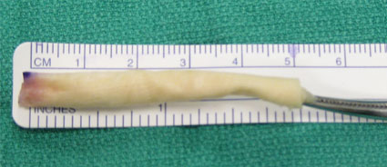
The graft used for allograft reconstruction is shown above. It is made from donor tissue from the iliotibial band. The graft is shaped and stitched before being placed into the hip joint.
Labral reconstruction is usually performed only when the labrum cannot be repaired. Sometimes the tissue is too damaged or worn down to hold stitches. In these cases, replacing the damaged tissue may be the best option. This decision is made during surgery after closely examining the labrum.
Fascia lata tissue from the iliotibial band is commonly used for reconstruction. Donor tissue used for surgery is carefully tested for disease.
Dr. Carreira has reported encouraging results in patients with severe labral damage. He also developed a fully arthroscopic reconstruction technique that he presented at an international conference and later published.
Treating other injuries around the hip joint at the same time as labrum surgery can help improve overall results.
All Arthroscropic Allograft Labral Reconstruction of the Hip
In spring 2014, Dr. Carreira presented his surgical technique using donor fascia lata tissue for irreparable labral tears at a meeting of the American Academy of Orthopedic Surgeons.
All Arthroscropic Allograft Labral Reconstruction of the Hip
Small Labrum Tissue Can Be a Candidate for Labrum Reconstruction in Revision Surgery


The above two images highlight a small labrum outlined by the red dots. As this particular labral tissue so small, it does not function like a normal labrum. In this case, the patient never developed a normal-sized labrum as they grew up.
Sometimes when this sort of small labral tissue is torn, it can be partially removed. That removal can result in a small labrum that is functionally deficient, in other words, non-functional. That is, the small labrum does not provide the support for regular movement of the hip. Where patients with small labrum experience persistent pain, they can be a candidate for labrum reconstruction in a revision surgery.
Photos of Allograft Labral Reconstruction of the Hip
Os Acetabuli with Too Little Labral Tissue
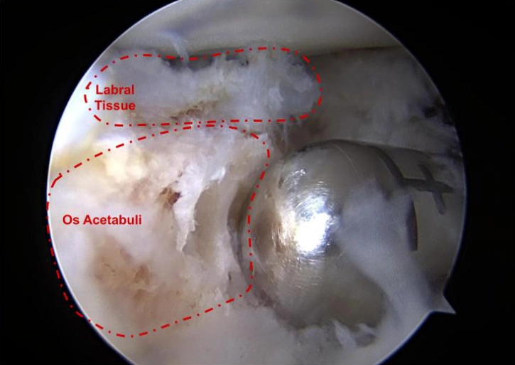
This image shows an os acetabuli with too little labral tissue as seen during hip arthroscopy. The os acetabuli should not be visible when labral tissue is normal, which is the reason for performing a labral reconstruction in this patient.
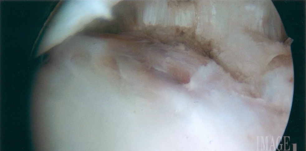
In this patient who underwent surgical treatment elsewhere, the labrum has been treated with electrocautery and does not appear healthy, with darkened and discolored appearance. This teenager underwent an allograft reconstruction.
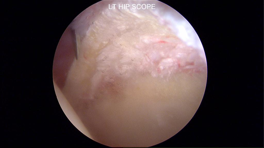
Taken during arthroscopic hip surgery, this picture demonstrates calcification of the labrum. The calcification is the white speckled appearance on the tissue itself. The calcification is a form of labrum degeneration. The issue was medically corrected via a reconstruction procedure.
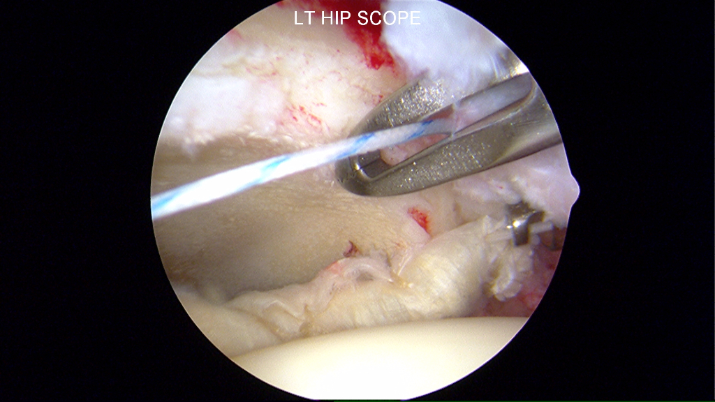
The tubular structure is the graft introduced into the joint. The structure is shown prior to refixation along the rim of acetabular bone.
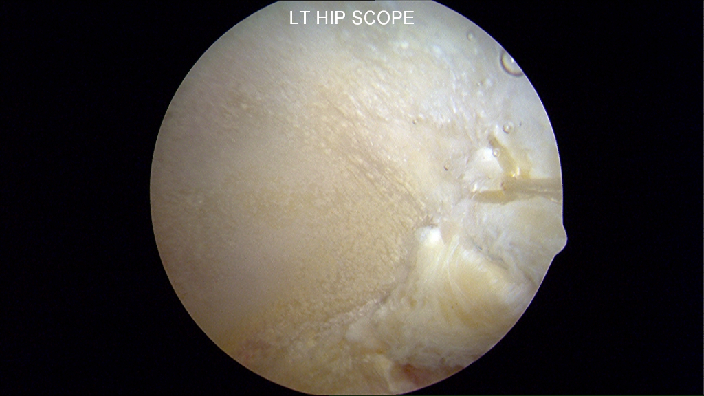
The tissue along the bottom right of this picture is the reconstructed allograft iliotibial band. The photo shows the tissue refixatation with suture.