
 Hip Problems + Treatments
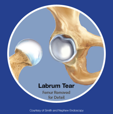
A hip labrum tear is a tear in the ring of cartilage around the hip socket. The hip joint is made of the top of the thigh bone fitting into the hip socket. The labrum helps deepen the socket, making the joint stronger and more stable. When the labrum tears, the hip joint may not work normally and may develop damage or arthritis over time.
Office Appointments and Telemedicine with Dr. Carreira

You can also book an office appointment or a telemedicine visit by calling Dr. Carreira’s office at 404-355-0743. Book now.
 What is the Labrum?
The labrum helps absorb shock, spread pressure, lubricate the joint, and keep the hip stable.
What is a Labral Tear?
The term labral tear includes many different kinds of damage to the labrum. Dr. Carreira compares the labrum to a rope tied to a boat dock. Over time, the rope may become worn out, frayed, partly torn, or almost completely torn. The same thing can happen to the labrum.
Labral Degeneration: Labral degeneration means the labrum has wear-and-tear damage over time. The tissue changes and becomes weaker with age. Injuries, hip impingement, loose joints, and hip dysplasia can also increase damage to the labrum.
Complexity of Tearing: Complex tears are tears that split the labrum in several directions and depths.
Hypoplastic: Hypoplastic means the labrum tissue is smaller than normal. Some patients naturally have less tissue, and others may have less tissue after a previous surgery.
What Causes An Acetabular Labrum Tear?
Hip labrum injuries can happen from repeated motion over time or from sudden injuries such as falls, twisting, or direct hits to the hip. Conditions like femoroacetabular impingement (FAI), Perthes disease, dysplasia, and slipped capital femoral epiphysis (SCFE) may also increase the risk of injury.
Diagnosis of Acetabular Labrum Tears
Signs and symptoms of a hip labrum tear include pain during movement, pain at night, stiffness, weakness, and reduced range of motion. Some patients also notice clicking, popping, grinding, or snapping in the hip.
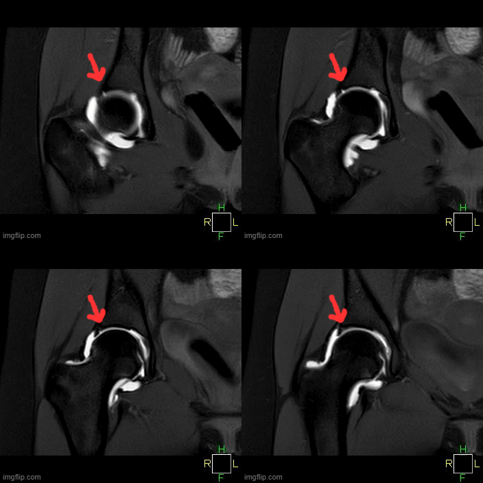
MRI can be useful for diagnosis of Labral Tears. Here we see images from the coronal view demonstrating a large labral tear. Note the elongated white signal at the arrow on multiple frames which shows the separation of the labrum from the acetabulum.
Treatment of Hip Labral Tears
Repair of a hip labrum tear means stitching the torn tissue back into place. Debridement means cleaning out damaged tissue and leaving healthy tissue behind. The labrum may be damaged in many different ways, including tearing, instability, and tissue breakdown. Studies increasingly show that repair often works better than debridement.
Several studies have shown high success rates for labral repair. Research comparing repair and debridement found that patients often had better results after repair surgery.
Labral Debridement
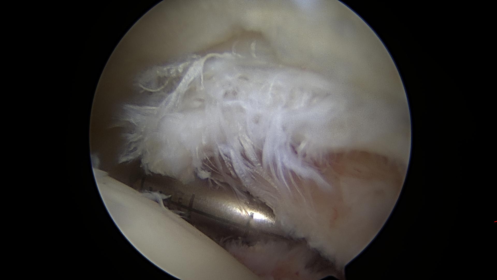
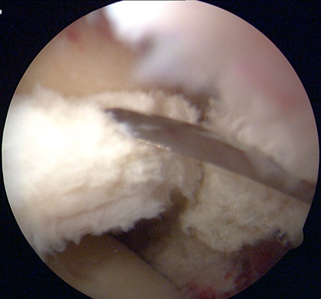
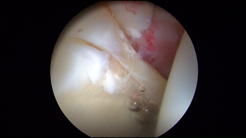
This is an example of a labral debridement. This patient had this injury towards the backside of the labrum in the joint and it simply needed to be smoothed out as demonstrated by the second picture. A “debridement” simply means removing tissue, and in this case, it previously looked like crab meat before it was smoothed out. In this patient, the front of the joint was repaired.
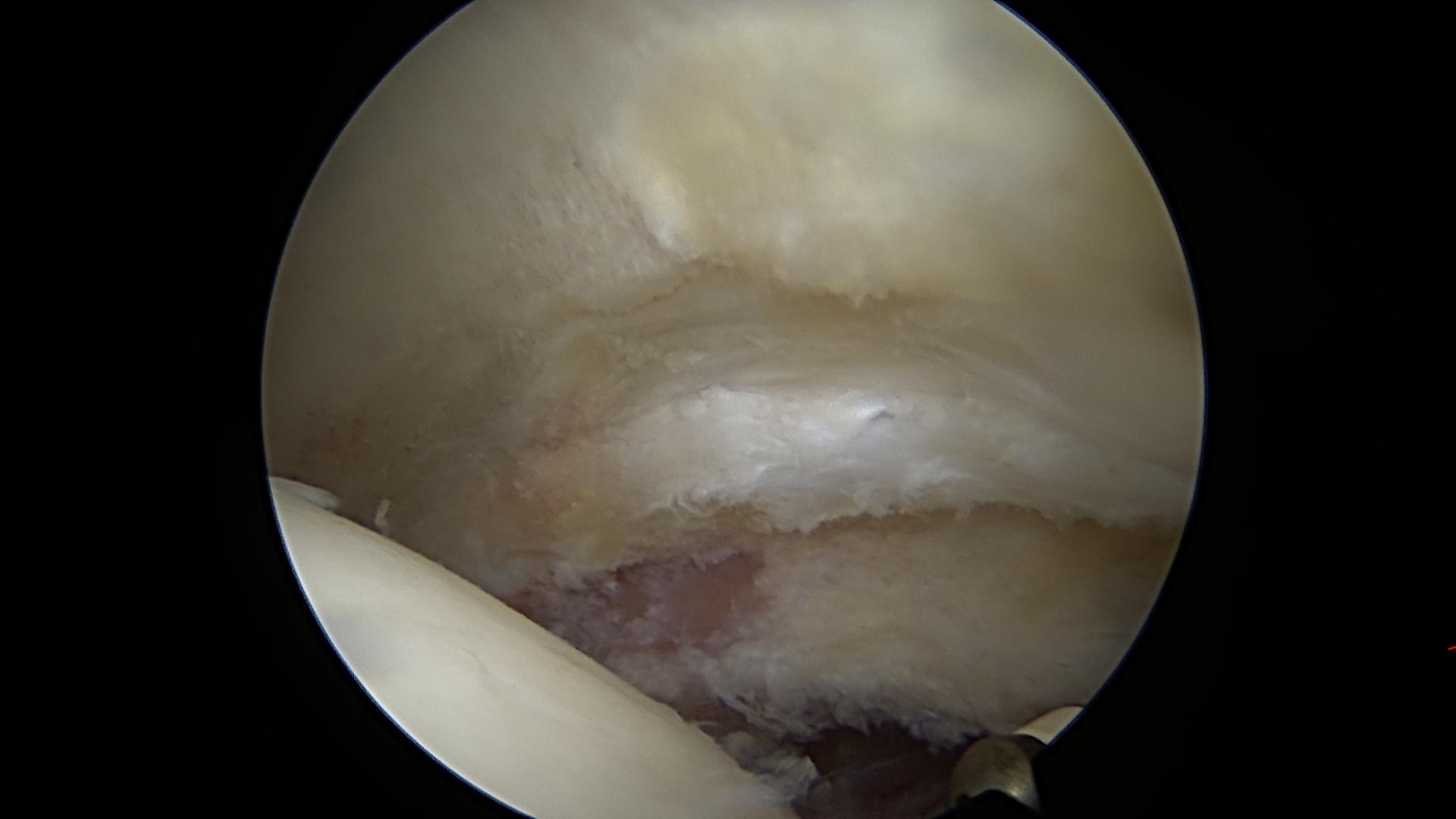
Hip Labrum Tear Video
Hip Labral Repair for Tear using Arthroscopy
Please note that there is no sound with the above video showing arthroscopic repair of a hip labrum tear.
In the arthroscopic video, the surgeon uses a probe to show unstable cartilage and labrum tissue inside the hip joint.
This video shows repair of cartilage during labrum repair surgery. Sutures are used to connect the cartilage and labrum together to create a smoother, more natural joint surface.
The above video is age-restricted on YouTube because it shows arthroscopic surgery.
Arthroscopic Repair of a Labral Tear with an Anchor and Suture Passer
This video demonstrates a labral repair. Anchors and stitches are used to hold the labrum in place.
Repair of Hyperplastic (Abnormally Large Diameter) Labral Tissue
The above video demonstrates repair of a healthy but unusually large labrum, called a “hyperplastic” labrum. Multiple anchors are used to stabilize the tissue.
All of the above videos are also available on Dr. Carreira’s YouTube channel.
Acetabular Labral Tears: Repair vs. Reconstruction
The choice between repair and reconstruction depends on the type and severity of the injury. This is still an area of debate among hip surgeons, and Dr. Carreira is a regular contributor to that academic conversation.
Researchers in the Multicenter Arthroscopy Study of the Hip continue studying the best ways to treat these injuries. Preserving and repairing the labrum is usually the main goal, and this treatment is Dr. Carreira’s main specialty.
Acetabular Labrum Repair with Four (4) Circumferential Sutures
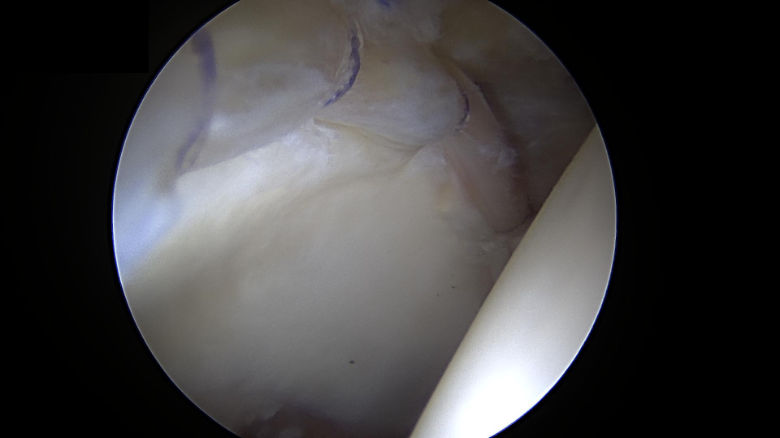
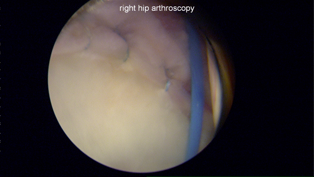
The above photo shows a hip labrum repair via four (4) circumferential sutures on the right hip of a 35-year-old woman.

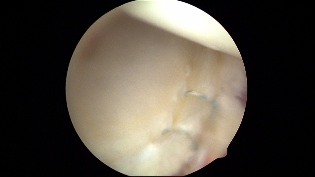
The above photo shows a hip labrum repair with four (4) anchors placed.
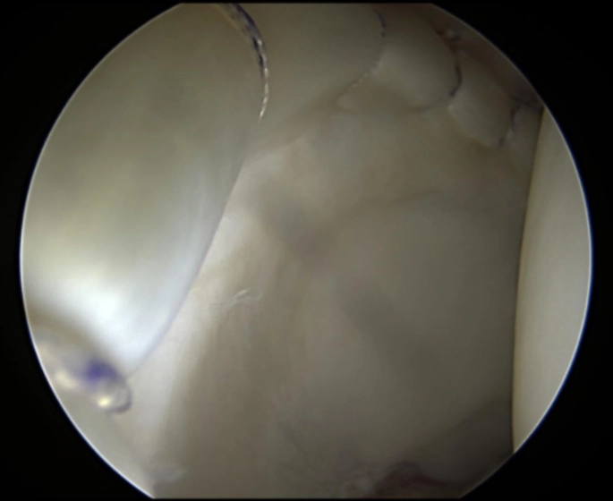
This arthroscopic image shows a healthy labrum repair using five anchors in a patient with a shallow hip socket and related early cartilage damage. This damage can be hard to detect in MRI or CT scans.
Acetabular Labral Reconstruction of the Hip – Addressing a Hip Labral Tear
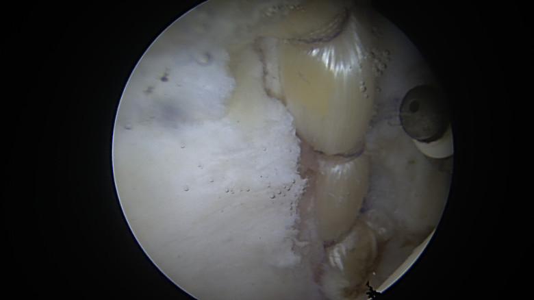
The above photo shows a labral reconstruction using donor tissue in a 29-year-old athlete.
Labral Reconstruction as a Revision Surgery
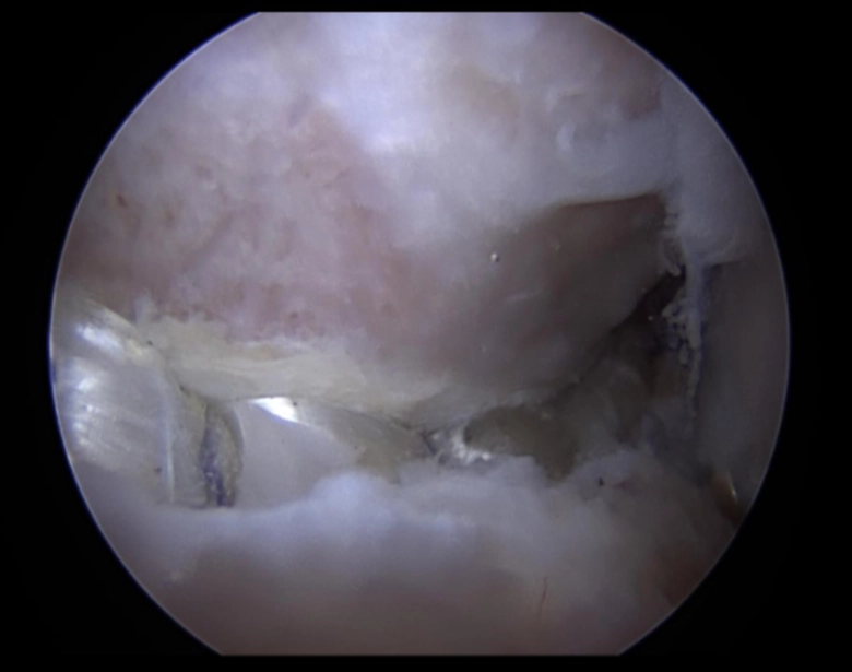
The image above shows a revision surgery where a new graft was used to rebuild the labrum. The labrum can be seen at the bottom of the screen, and the femoral head is at the top of the image. The femoral head had been previously trimmed in impingement treatment. With the graft in place, impingement testing is performed by moving the hip in various ways to ensure it moves smoothly.
Labrum Damage Including Degeneration and Core Bruising
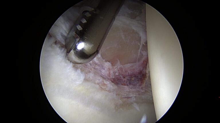
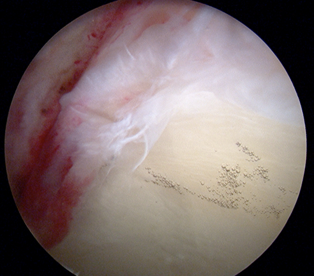
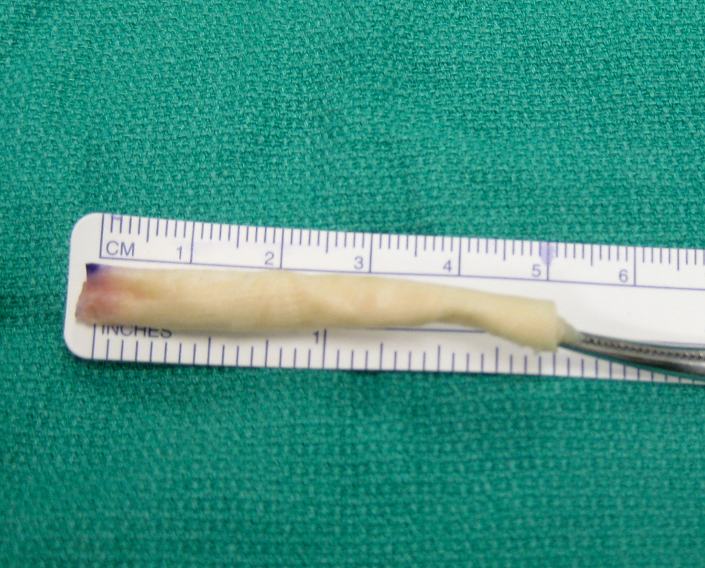
The two images below show damage to the labrum including degeneration and bruising. The second picture shows the labral tissue removed, demonstrating the inflammation in the center of the tissue in red.



Additional Hip Labral Issues and Surgical Procedures
Hip Labrum Repair with Five (5) Anchors
This arthroscopic video shows a labral repair using five anchors. The tissue appears healthy with only a simple detachment and little degeneration. The size of the labrum also appears to be normal.
The MASH– Multicenter Arthroscopy Study of the Hip- Study Group has published research on the relationship between different kinds of labrum injury and the outcomes of treatment. Labrum injury is more complex than simply a tear or not.
Labral degeneration can make repair surgery less successful, although many patients still improve after surgery.
Labral Tear with Articular Cartilage Flap
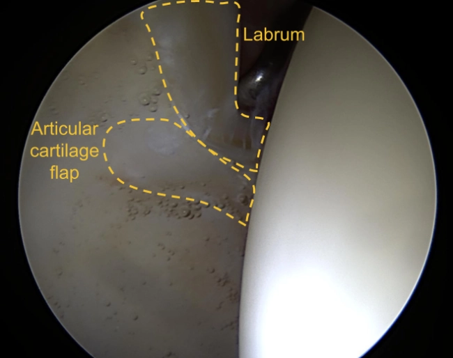
The image above shows the front of the acetabulum with a labral tear and a flap of articular cartilage. That tear and flap of cartilage was causing a patient pain and discomfort. To address the issue, the flap was trimmed down and the labrum was repaired, all via arthroscopic surgery. Following the procedure, the patient experienced a fuller return to motion with no more pain.
Burr Used to Stimulating the Bone for Healing of the Labrum
A burr tool is used to prepare the bone and create a better healing environment before the labrum is repaired.
Chondrolabral Repair via Arthroscopic Surgery of the Hip: Repair of the Articular Cartilage
Chondrolabral Dysfunction
In the video below, the labrum and cartilage move too much because they are unstable.
During surgery, anchors and sutures were used to stabilize the tissue, and the patient had an excellent result.
The video also shows inflammation in the joint lining, called synovitis, as noted by the reddened appearance along the top of the screen.
Chondrolabral Repair via Arthroscopic Surgery of the Hip: Unstable Flap of Cartilage
All Arthroscropic Allograft Labral Reconstruction of the Hip
This video is age-restricted on YouTube because it shows surgery.
Hip Arthroscopy Placement Suture Anchor
YouTube automatically placed an age restriction on the video because it shows an arthroscopic surgical procedure.
Complex Labral Injuries
Dr. Dominic Carreira and his team of hip specialists in the MASH Study Group have focused much of their research on the complexities and subtletiesof labral injuries. As such, Dr. Carreira and his colleagues are uniquely positioned to highlight and discuss these potentially life-changing injuries. Through the content in this section, Dr. Carreira will make it clear that a labral tear can often prove a complicated problem – much more than a simple tearing.
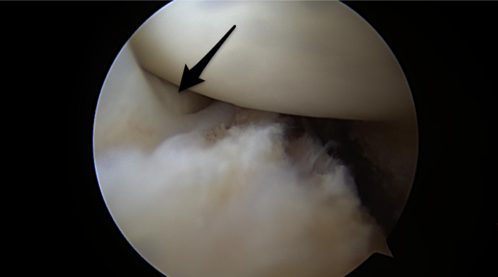
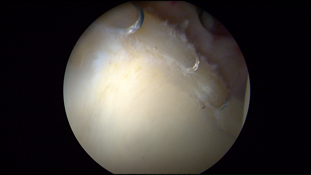
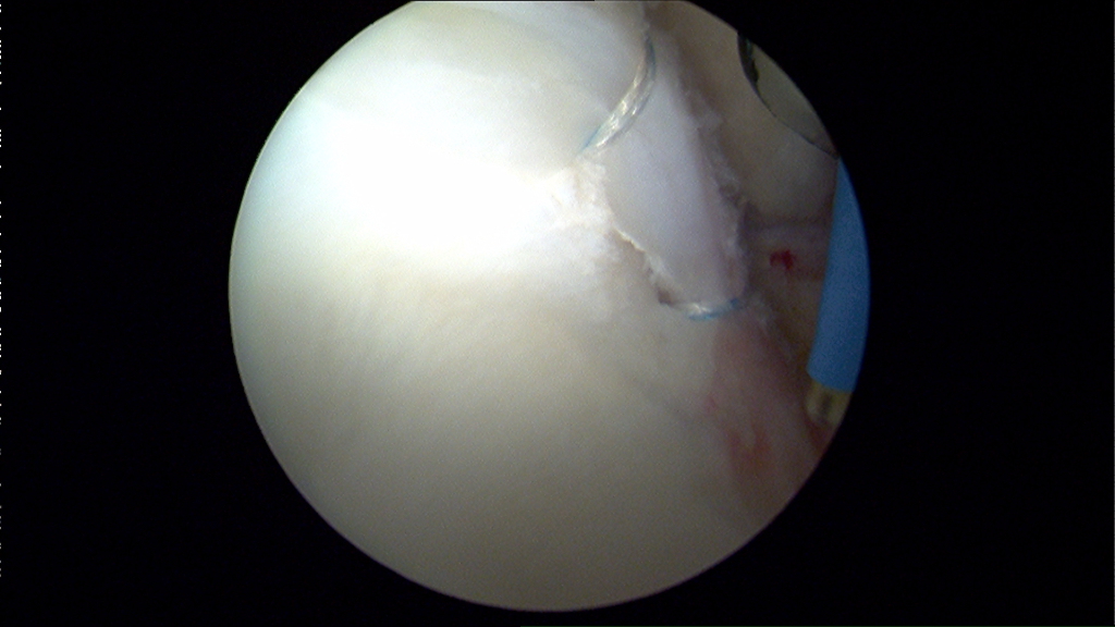
The gallery below of arthroscopic images detail picture a labrum which is not positioned well. As a result of this poor positioning, the patient’s labrum does not have a good suction seal. That lack of a proper seal means that the patient’s labrum cannot function as it should. The articular cartilage (arrow shown) should not be visible from this view outside of the hip joint. In the latter two images, the labrum is moved into to the correct position. Because of this corrective repositioning, the patient’s articular cartilage is no longer visible.

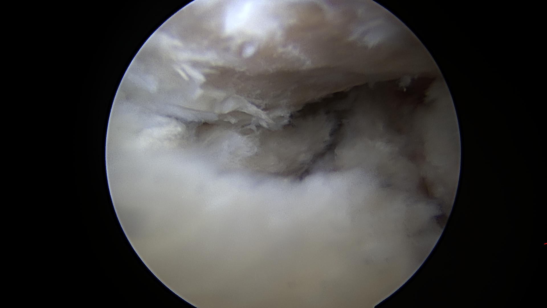
Labral Exhibiting Tearing and Degeneration

A labral with severe complexity of tearing with degeneration.
Healthy Labrum Following Arthroscopic Repair

This labral repair demonstrates very healthy labral tissue, typical for a young patient who has had symptoms for a short period of time. The labrum was repaired via arthroscopic surgery.
Photos of Hip Labrum Tear (Acetabular Labrum Tear)
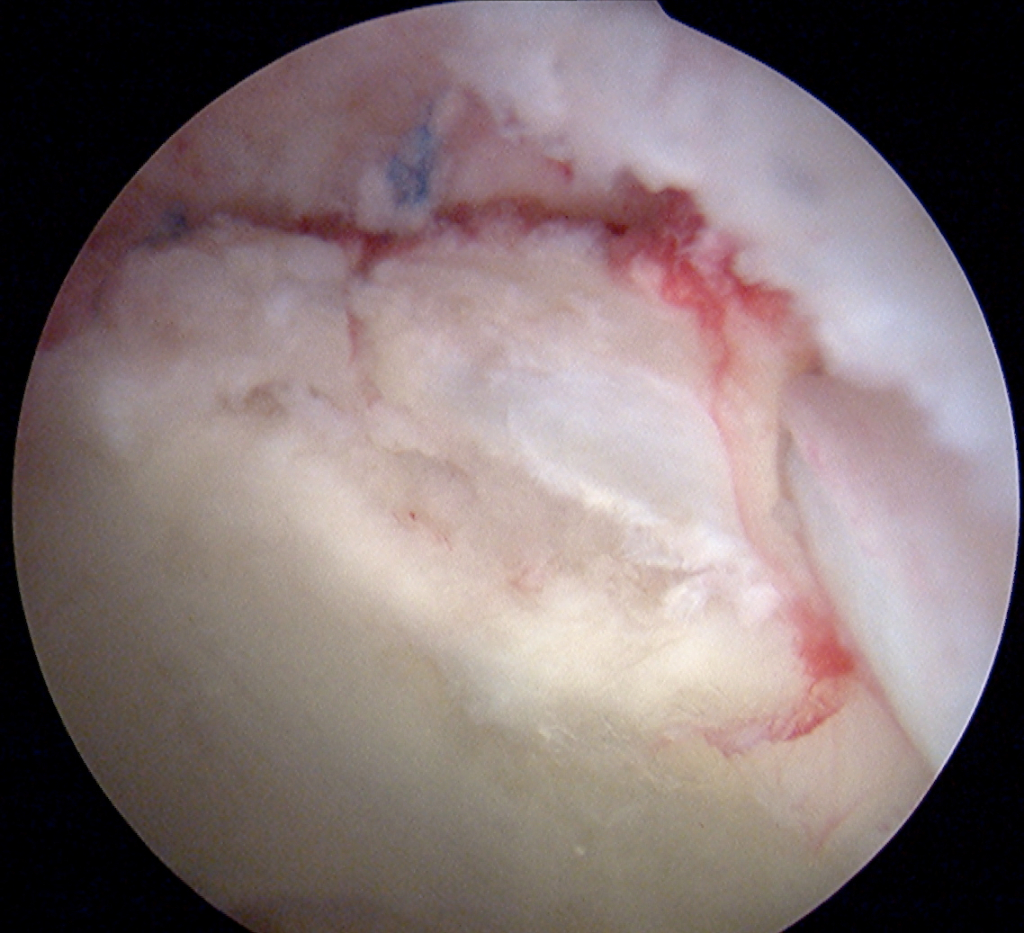
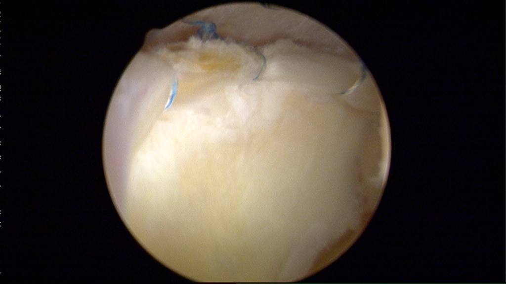
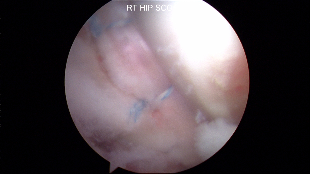
Photos of Hip Labral Repair