
Some patients have an extra bone in the foot called an accessory navicular. In certain patients, this bone can become very painful. The typical presentation for these patients is pain along the inside and back of the foot. The accessory navicular is a protruding abnormal bony prominence, and in some patients may never become painful. Treatment is done only for those patients who have pain. The treatments include activity modification, orthotic inserts to support the arch and offload this area, medications such as nonsteroidal anti-inflammatory’s (nsaids), and placement into a boot or cast.
Office Appointments and Telemedicine with Dr. Carreira
You can also book an office appointment or a telemedicine visit by calling Dr. Carreira’s office at 404-355-0743. Book now.
Kidner Procedure
If these non-operative measures fail, the option of surgical treatment can be performed. The terms used to describe the procedure include removal or excision of the bone that never fused normally in development and/or removal of the prominence of bone of the navicular.
The surgery is also called the Kidner procedure. If there is a flatfoot associated with the os navicular, the flatfoot can also be corrected to help restore alignment and offload the reconstruction. This consists of a posterior tibial tendon reattachment. The posterior tibial tendon normally inserts on this bone so a reconstruction is necessary when the bone is removed.
Typically the rehabilitation course consists of non-weight bearing for two weeks in a short leg cast. This is followed by CAM boot immobilization and full weight-bearing for an additional 4 weeks.
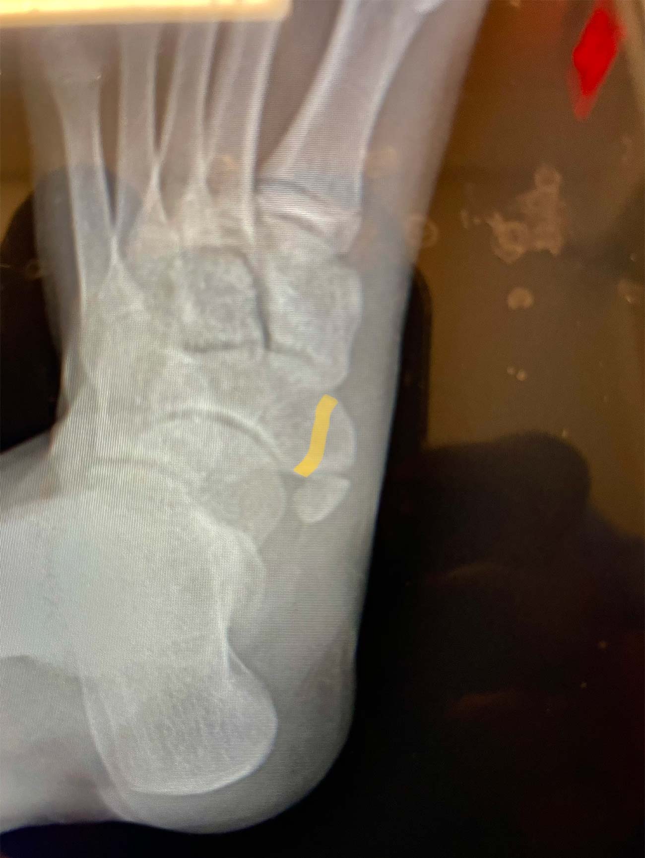
This picture above shows a yellow line which depicts the portion of the navicular body which is removed to create a smooth inner border to the foot. The os navicular bone itself, seen here as an oval round bone, is also removed at the same time.
Before and After X-Rays of Os Navicular
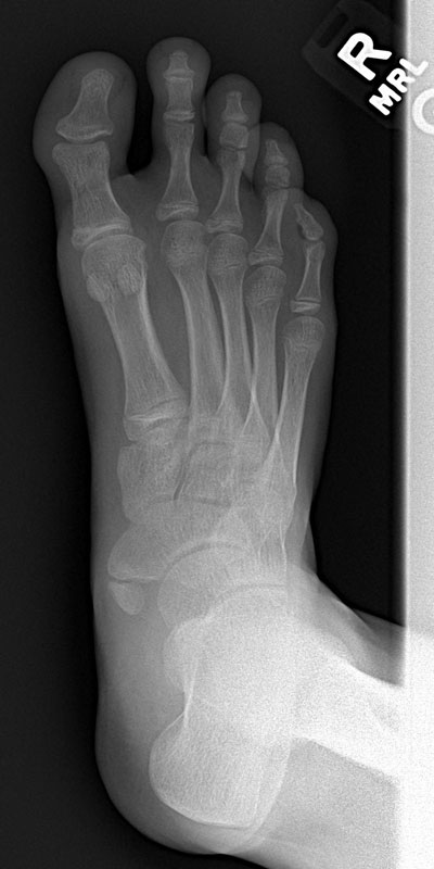
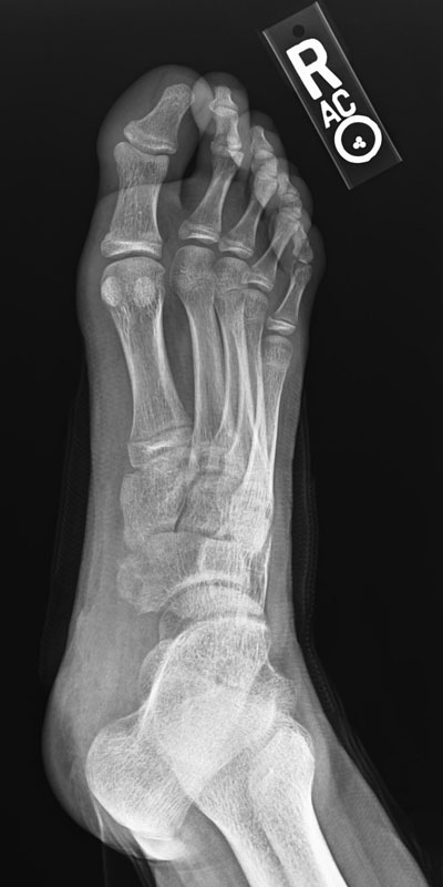


This post-op x-rays images above demonstrate the resection and re-creation of a smooth and flat inner border of the foot. The first x-ray on the left is taken before the procedure. The second x-ray, on the right, is taken after successful completion of the procedure.