
Benign synovial diseases found in the hip include pigmented villonodular synovitis (PVNS) and synovial chondromatosis (SC). Although these synovial disorders are infrequently encountered, failure to recognize and correct them in a timely fashion may accelerate the progression of joint degeneration and cartilage damage.
Office Appointments and Telemedicine with Dr. Carreira
You can also book an office appointment or a telemedicine visit by calling Dr. Carreira’s office at 404-355-0743. Book now.
What is Pigmented Villonodular Synovitis (PVNS)?
Pigmented Villonodular Synovitis (PVNS) is a joint disease of the synovium, or joint lining. It is characterized by inflammation and overgrowth of the synovium and it typically affects the hip (or ankle.) The overgrowth and inflammation harms the joint, leading to early joint damage. Typically patients complain of pain and restricted movement.

The above nodule of diseased PVNS was removed during a hip arthroscopy procedure.
PVNS is idiopathic, i.e. it occurs without any specific cause. There is no genetic or hereditary association, and it is not related to activity levels.
Video of Pigmented Villonodular Synovitis (PVNS)
Surgical Treatment Options of Pigmented Villonodular Synovitis (PVNS)
Surgery is typically recommended as a treatment for PVNS, with the type of surgery dictated by the extent of damage to the joint. In cases where significant degeneration has occurred and the damage is more advanced, a joint replacement is recommendable in the hip. In cases of minimal joint damage, an arthroscopic procedure may be performed to remove as much of the diseased synovium as possible. Even with surgery, the PVNS may recur and require additional treatments including repeat surgery or radiation therapy.
Dr Carreira has published an article on this specific topic. The article, A systematic literature review of synovial chondromatosis and pigmented villonodular synovitis of the hip, was published in the journal THE PHYSICIAN AND SPORTSMEDICINE in 2016. The review identified and systematically summarized 12 studies reporting population characteristics, imaging modalities, treatments, and outcomes for PVNS and synovial chondromatosis (SC) in the hip. Major findings include the importance of early diagnosis and surgical intervention to achieve satisfactory outcomes, and the lack of consensus regarding the effectiveness of open versus arthroscopic surgical debridement.
For more information about PVNS or to schedule an appointment, please contact Dr. Carreira directly.
Photos of Surgical Treatment of Pigmented Villonodular Synovitis (PVNS)
The following three images of PVNS show it before surgical debridement and after an arthroscopic surgical debridement/clean out, as well as a typical nodule removed during such a procedure.
PVNS – Before Debridement Clean Out
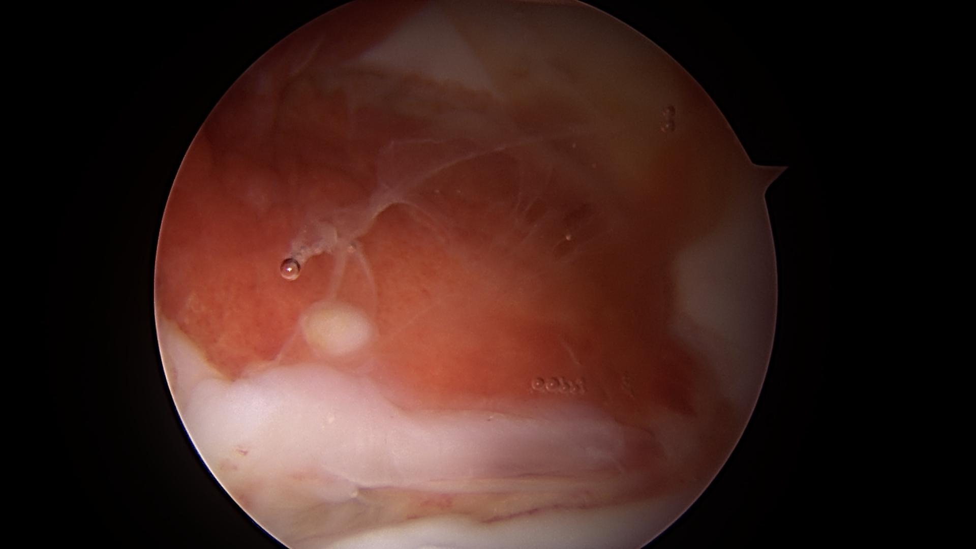
Arthroscopic view of severe synovial inflammation, or synovitis, related to pigmented Villo nodular synovitis (pvns). These lesions appear suspicious at the time of arthroscopy, because of their more characteristic, yellowish color and nodular appearance compared to standard synovitis. The diagnosis is confirmed with biopsy.
PVNS – After Debridement Clean Out
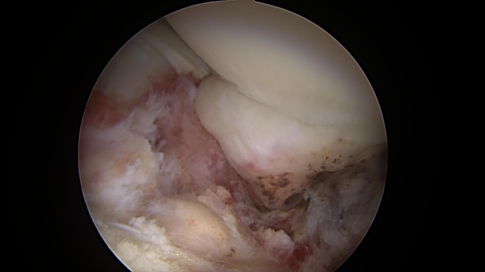
View inside the hip following clean out or debridement of the PVNS. The treatment is to remove the abnormal synovium as extensively as possible, including in both the central and peripheral compartments of the hip
PVNS – Removed Nodule
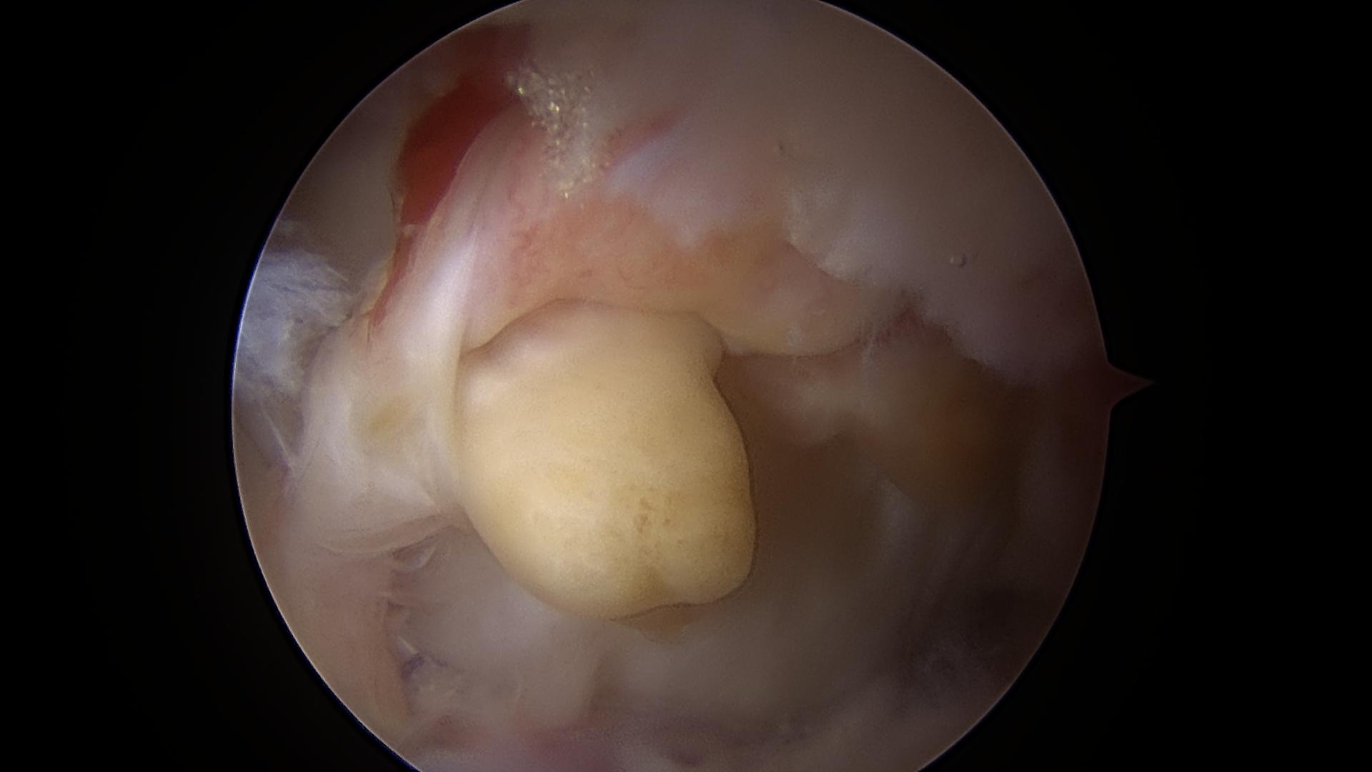
This is a characteristic nodule of PVNS which was removed at the time of hip arthroscopy.
Photos of Pigmented Villonodular Synovitis (PVNS)


