
 Hip Problems + Treatments
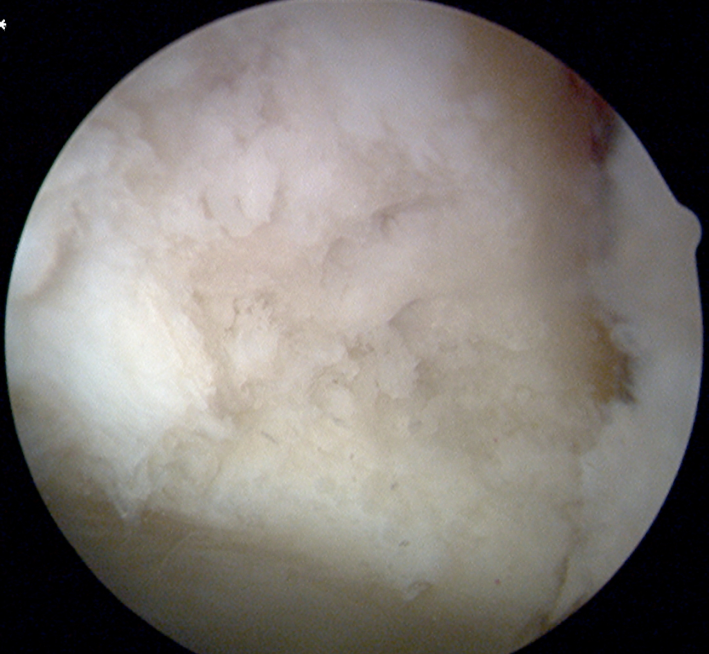
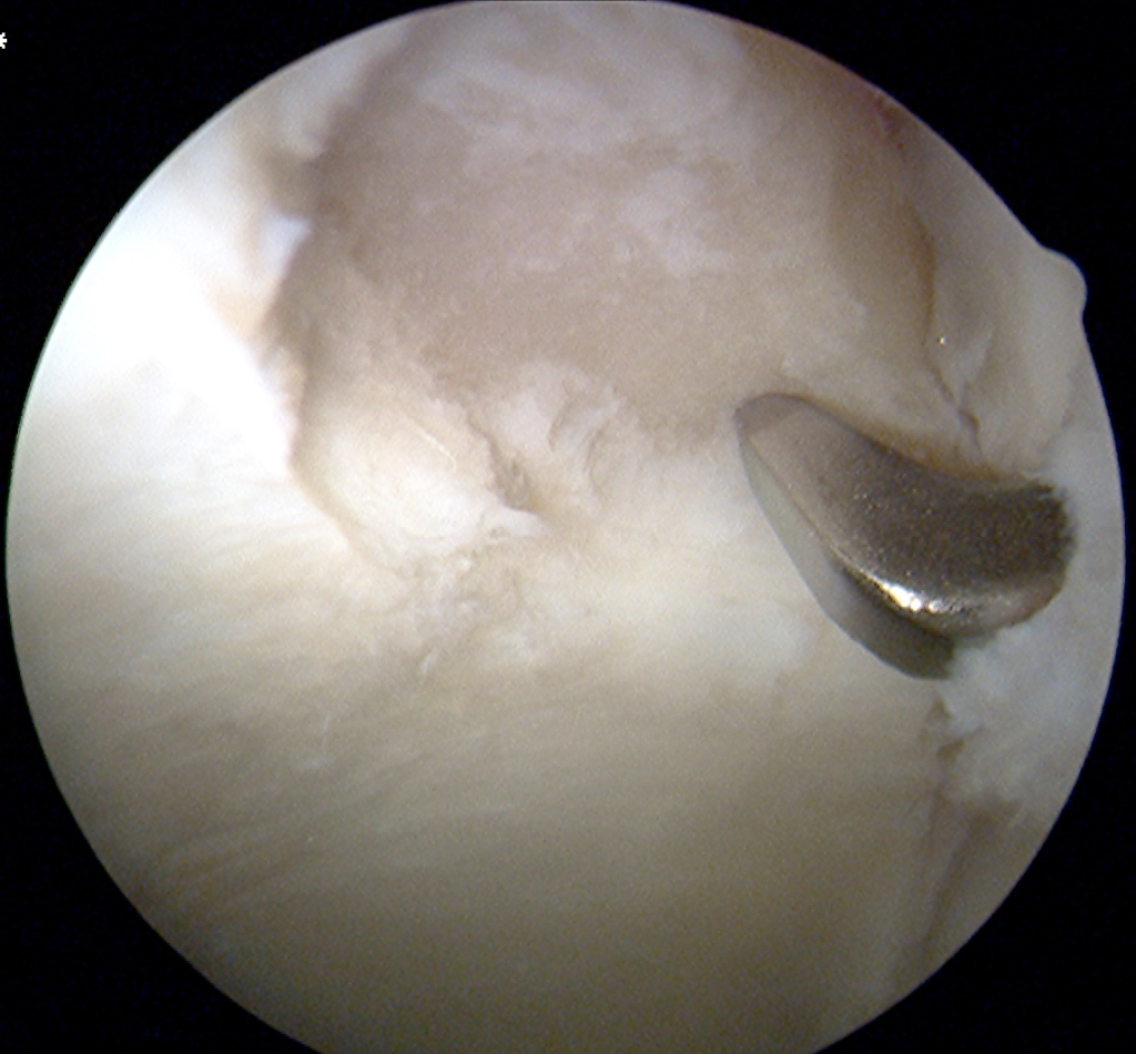
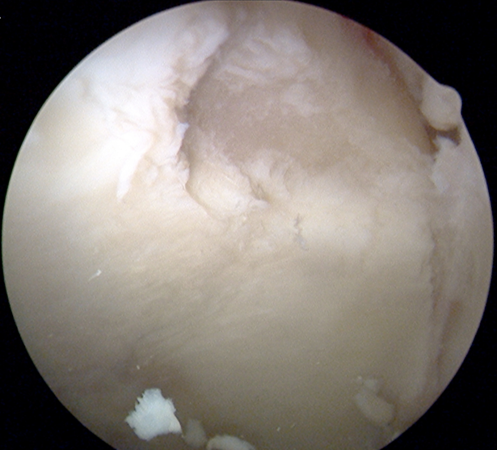
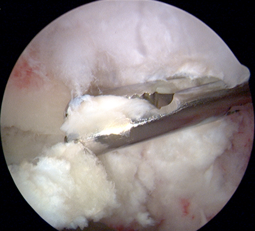
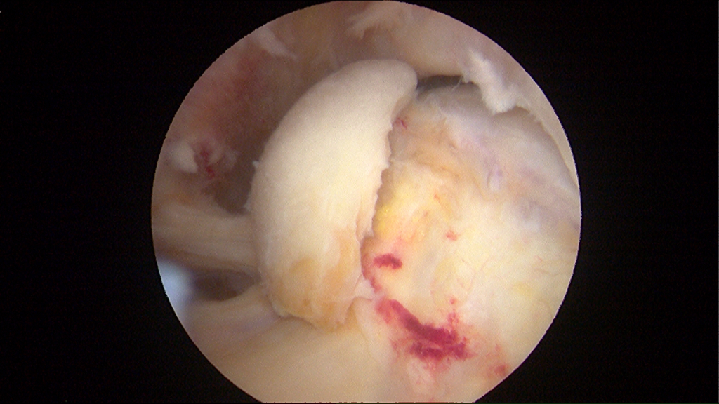
Focal cartilage damage is a small area of injury to the cartilage lining of the hip joint. Damaged cartilage can cause pain, stiffness, and decreased movement and function.
Office Appointments and Telemedicine with Dr. Carreira

You can also book an office appointment or a telemedicine visit by calling Dr. Carreira’s office at 404-355-0743. Book now.
What Are the Causes of Articular (Hip) Cartilage Injury?
There are many causes of cartilage damage, including genetics, aging, overuse injuries, and obesity, to name a few. Healthy cartilage cushions the joints and allows for smooth, gliding movements. Your doctor will evaluate you and help make this diagnosis.
Treatment of Articular Hip Cartilage Defects
One treatment option is called an “arthroscopic microfracture procedure”. An “arthroscopic” procedure means that small incisions are made to insert a camera and surgical tools, leading to quicker recovery times. During an arthroscopic microfracture procedure, damaged cartilage is removed, and tiny holes are made in the bone underneath to help stimulate the growth of new cartilage tissue.
Over several weeks, the body creates new cartilage-like tissue in the damaged area, which may reduce or eliminate pain. This treatment works best for smaller areas of damage and is successful in about 85% of patients. It is not recommended for patients with widespread arthritis.
Hip Microfracture
This arthroscopic procedure demonstrates the microfracture technique. This technique is used for joint cartilage defects and is used in multiple joints. The surgery usually requires limited weight bearing with crutches or a walker for at least 6 weeks after surgery. Continuous passive motion (CPM) machines may also be prescribed for recovery.
Additional videos are available for review on Dr. Carreira’s Youtube channel.
Wave Sign or Carpet Phenomenon in Labrum and Articular Cartilage Injury
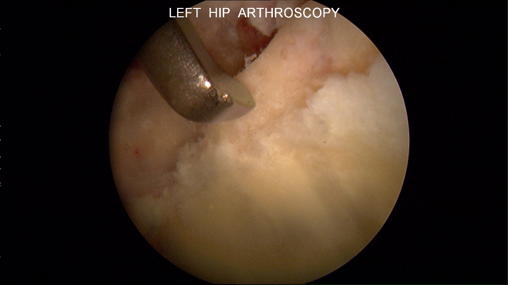
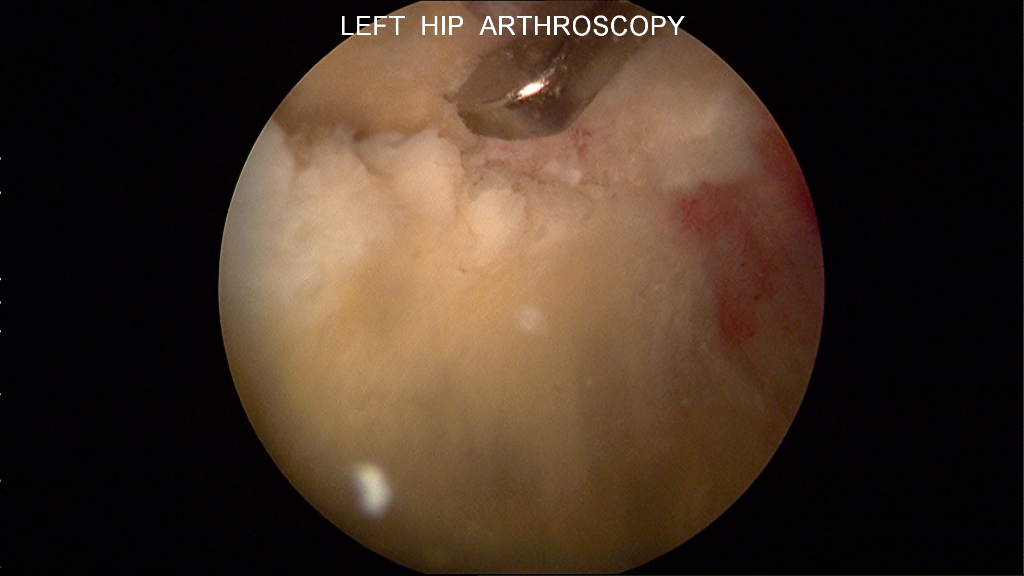
The above video demonstrates a common pattern of labrum and cartilage injury called the “wave sign” or “carpet phenomenon.” The damaged cartilage (seen in the middle) is no longer firmly attached to the bone underneath it, unlike the healthy cartilage nearby (seen on the left).
Photo of Femoral Head Chondrosis
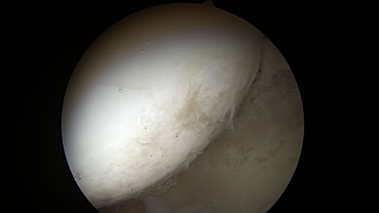
Femoral head chondrosis (cartilage damage) that is less than 50% of cartilage depth as noted during a hip arthroscopy procedure.
Severe Cam-Type Hip Impingement
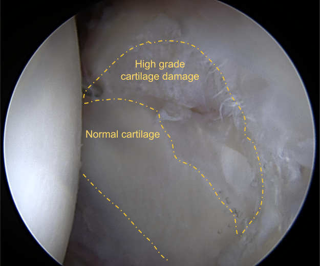
The above image shows the hip of a patient in their early 20s with severe cam-type hip impingement. That impingement has led to progressive cartilage damage after years of being left untreated. The severity of cam impingement is important in the prognosis, with such impingement a notable concern for the mobility and function of the patient’s hip.
Hip impingement like what is shown in the above graphic typically results from athletic activities during adolescence, with an increased risk in high-impact activities or contact sports. Hip arthroscopy to treat severe impingement is typically recommended when the impingement is as severe as it was in this patient. Such impingement can lead to cartilage damage over time.
Dr. Carreira treated the patient by trimming the excess bone during arthroscopic surgery of the hip, in a procedure called a femoroplasty.
Photo of Central Osteophytes (Arthritic Boney Spurs) and more
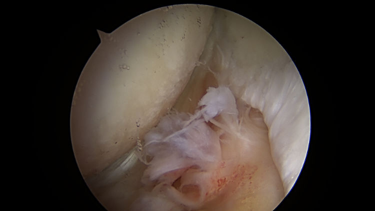
Central osteophytes (arthritic boney spurs), femoral head chondrosis (damage of the cartilage on the femoral side of the hip joint) and tear of Ligamentum teres noted during hip arthroscopy in a 47-year-old male. These findings of intra-articular damage place this patient at greater risk of needing a hip replacement in the future.
Photos of Limited Arthritis / Focal (Hip) Cartilage Damage