What is Pigmented Villonodular Synovitis (PVNS)?
Pigmented Villonodular Synovitis (PVNS) is a joint disease of the synovium, or joint lining. It is characterized by inflammation and overgrowth of the synovium and it can effect many joints, including the ankle and foot. The overgrowth and inflammation harms the joint, leading to early joint damage. Typically patients complain of pain and restricted movement.
Office Appointments and Telemedicine with Dr. Carreira

You can also book an office appointment or a telemedicine visit by calling Dr. Carreira’s office at 404-355-0743. Book now.

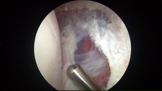
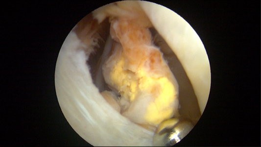
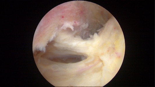
The above photos shows the typical appearance of PVNS. There is a golden brown appearance with areas of yellow very friable tissue.
PVNS is idiopathic, i.e., it occurs without any specific cause. There is no genetic or hereditary association, and it is not related to activity levels. The diagnosis is confirmed at the time of surgery, and there are MRI findings that may suggest its presence and provide a high suspicion of it being present. The MRI can show clusters of thickening of synovium not only around the joint itself surrounding the capsule but the PVNS may extend beyond the boundaries of the joint itself as the disease progresses.
Video of Pigmented Villonodular Synovitis (PVNS)
Surgical Treatment Options for Video of Pigmented Villonodular Synovitis (PVNS)
Surgery is typically recommended as a treatment for PVNS, with the type of surgery dictated by the extent of damage to the joint. In cases where significant degeneration has occurred and the damage is more advanced, a joint replacement or fusion is recommendable. In cases of minimal joint damage, an arthroscopic procedure may be performed to remove as much of the diseased synovium as possible. Even with surgery, the PVNS may recur and require additional treatments including repeat surgery or radiation therapy.
For more information about PVNS or to schedule an appointment, please contact Dr. Carreira directly.
Photos of Pigmented Villonodular Synovitis (PVNS)
Treatment of Abnormal Synovium in PVNS
As noted above, surgery is the most common treatment option for Pigmented Villondular Synovitis (PVNS) of the ankle. In the images and video that follow, Dr. Dominic Carreira shares an example of a successful arthroscopic procedure wherein he removed abnormal synovium from a patient.
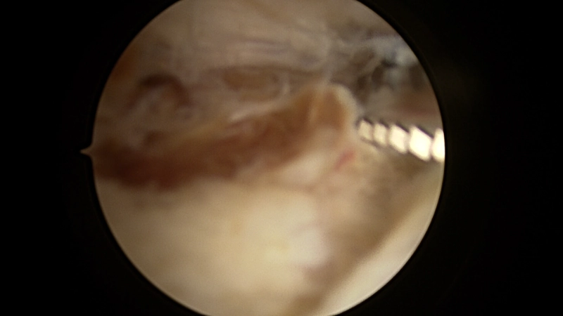
This is the posterior ankle joint line demonstrating the abnormal synovium in PVNS. Note the golden brownish discoloration which is the hallmark of this disease.
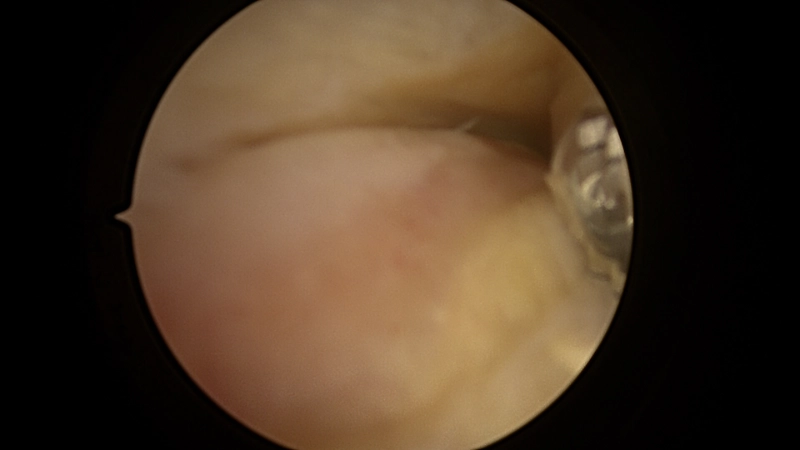
The subtalar joint seen from a posterior approach. There is a triangle in the right upper portion of the screenshot showing the golden brownish discoloration typical of PVNS. All of this discolored tissue was removed with a shaver.
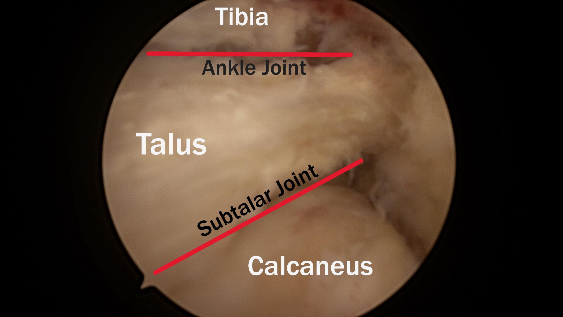
There are two (red) lines showing the ankle and subtalar joints as viewed arthroscopically from a posterior approach. No residual PVNS is noted when looking at both of these joints after the successful removal of the abnormal tissue via arthroscopic surgery.
In this video, the shaver being used to remove the PVNS from the back of the ankle joint. The golden brownish tissue is removed, along with the reddened tissue, which is inflamed because of the PVNS. An extensive search for abnormal tissue is done throughout the entire joint.


